AI-designed peptides: what new lab tests show, and what they don’t
News
Four 2026 papers test computer-designed peptides and prediction models at the bench — all lab and computational work, with no human data.
Peptide design has shifted from hand-tuning sequences to asking software what to make next. Four papers published in 2026 show where that shift stands: a de novo designed peptide that copies how a broadly neutralizing antibody grips an influenza surface protein [1], a model that predicts how a peptide’s folding changes with its chemical surroundings [3], a large prospective test of how well deep-learning co-folding tools actually place ligands [2], and an interpretable framework for navigating astronomically large peptide design spaces [4]. All of it is computational and laboratory work. None of these studies involved people.
Key takeaways
- A hotspot-guided computational design produced short peptides that bound influenza A hemagglutinin in the same way two broadly neutralizing antibodies do, and blocked the low-pH shape change that drives membrane fusion in vitro [1].
- An environment-conditioned model trained on more than 1,500 peptides and more than 5,500 peptide-environment measurements predicted how secondary structure redistributes between water, co-solvent, and membrane-mimicking conditions [3].
- Tested prospectively on 489 evaluable ligand complexes determined after their training cutoffs, three co-folding tools each reproduced more than half of poses to better than 2 Å, but missed the protein conformational rearrangements that accompany some binding events [2].
- These are design and prediction studies in silico and in vitro. No clinical outcomes, no safety data, and no human testing are reported in any of the four papers [1, 2, 3, 4].
Copying an antibody’s grip with a much smaller molecule
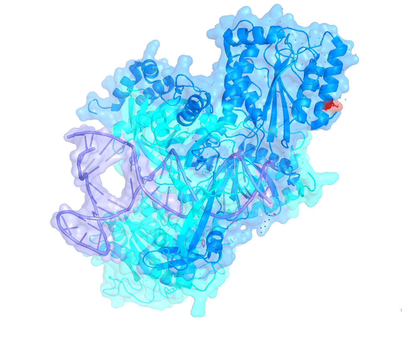
The first study is a computational design project followed by laboratory synthesis and structural work — no cells beyond in vitro assays, no animals, no people [1]. The idea is “hotspot-centric” design: take the handful of amino acid residues that do most of the binding work at an antibody-antigen interface, strip them out of the antibody, and graft those positions onto a much smaller scaffold that can hold them in the right geometry.
The authors used crystal structures of two broadly neutralizing antibodies, FI6v3 and CR9114, bound to influenza A hemagglutinin as their templates [1]. They selected hotspot residues from those antibodies and anchored them onto known peptide scaffolds of 35 amino acids or fewer drawn from the Protein Data Bank [1]. The top-scoring designs were then synthesized, folded, tested in vitro, and characterized structurally in complex with hemagglutinin [1].
The reported result is both structural and functional mimicry: the designed peptides recapitulated the binding mode of the parent antibodies on hemagglutinin, and acted as inhibitors of the low-pH-dependent conformational transitions that hemagglutinin undergoes to drive membrane fusion [1]. The authors frame this as extending a strategy previously applied to proteins and d-peptides into l-peptide scaffolds [1].
The limits are substantial and worth stating plainly. This is a demonstration that the design method works, assessed with binding and conformational assays and structures of the complexes — not an efficacy study. The abstract reports no animal infection experiments, no measures of stability or distribution in a living organism, and no human data [1]. Inhibiting a conformational change in a purified protein is several long steps away from affecting an infection in a person, and nothing in this paper addresses those steps.
Predicting shape-shifting, not just one shape
Most structure prediction returns a single model. Short peptides are a poor fit for that assumption, because many of them are only loosely folded in water and adopt helices or other structures when they meet a membrane or a different solvent. The third paper asks whether that responsiveness is itself learnable [3]. It is currently a preprint on bioRxiv, so it has not completed journal peer review [3].
The authors assembled an experimental dataset of more than 1,500 peptides and more than 5,500 peptide-environment observations spanning aqueous, co-solvent, and membrane-mimicking conditions, with secondary structure read out by circular dichroism [3]. Their framework, ApexFold, combines sequence representations with physicochemical descriptors of the surrounding medium to predict the fractions of α-helical, β-like, and unstructured conformations [3].
The test that matters most here is on two panels collected later and excluded from model development [3]. On those, the model captured both the direction and the magnitude of environment-induced structural redistribution, and the peptide-specific degree of plasticity, outperforming solvent-agnostic and composition-based baselines [3]. The authors point out that static structural references, which return one conformation, cannot represent condition-dependent response profiles at all [3].
What this does not show: circular-dichroism-derived secondary structure fractions are a coarse summary, not an atomic structure, and membrane-mimicking buffers are not membranes in a cell. The study is about predicting a physical property of molecules in solution [3]. It makes no claims about biological activity in cells, animals, or people.
A prospective reality check for co-folding tools
The second paper is the most direct audit in this group [2]. Deep-learning co-folding methods predict a protein and its bound ligand together, but evaluating them is hard because test molecules are often already in the training data. The authors handled this by testing predictions against 551 ligands bound to the SARS-CoV-2 NSP3 macrodomain (Mac1) whose structures were determined after the tools’ training cutoff dates; 489 were of sufficient quality to evaluate [2].
AlphaFold3, Boltz-2, and Chai-1 each reproduced more than 50% of the ligand poses to better than 2 Å root-mean-square deviation from experiment [2]. But a key hope for co-folding — that it would capture protein shape changes that stabilize binding — was not borne out: common rearrangements, including a peptide flip and a large loop opening, were not recapitulated [2]. On ranking, pose-prediction confidence from AlphaFold3 and Chai-1 tracked experimental potency weakly but significantly, DOCK3.7 energies correlated only weakly, and Boltz-2’s affinity predictions correlated most strongly, beating a baseline predictor on mean absolute error after calibration [2].
The authors also tested whether co-folding scores could rescore docking hit lists across hundreds of molecules experimentally tested against AmpC β-lactamase and the dopamine D4 and σ2 receptors [2]. AlphaFold3 pose confidence did not separate true ligands from high-scoring false positives as well as docking scores or Boltz-2 affinity predictions [2]. Their conclusion is modest and useful: physics-based and deep-learning scores correlate independently enough that combining them may help prioritize hits [2]. Note that these are small molecules against protein targets, not peptides — the relevance to peptide design is methodological.
Searching design spaces too large to enumerate
The fourth paper addresses a practical bottleneck. Adding noncanonical amino acids expands peptide chemical space beyond 10^50 candidates, which breaks conventional structure-activity models in three ways: they transfer poorly from natural residues to new building blocks, they are hard to interpret, and they do not scale [4].
PepOpt, the framework described, unifies molecule-based and sequence-based representations inside linear models and uses dynamic programming to search those spaces without enumerating sequences [4]. The reported advantages are accuracy with interpretability, inference across diverse noncanonical residues including ones absent from training whose constituent substructures appear in the training data, and decomposition of each prediction into residue-specific contributions [4]. The authors also note it handles multi-parameter optimization — potency alongside developability properties such as stability and solubility — without special modification [4].
This is a methods paper, positioned as a platform for prediction and ideation [4]. The abstract reports no prospective wet-lab validation campaign, and no biological testing in cells, animals, or people [4].
What we still don’t know
- Whether any of the designed influenza-targeting peptides do anything in a living organism. The study reports in vitro inhibition of a conformational transition and structures of the complexes, not infection outcomes [1].
- How these peptides behave over time in a biological setting. Degradation, clearance, and distribution are not addressed in any of the four papers [1, 2, 3, 4].
- Whether predicted structural plasticity translates into predicted function. ApexFold predicts secondary structure fractions under defined conditions, not activity [3].
- Why co-folding tools miss conformational rearrangements, and whether that can be fixed. The authors document the gap on one well-characterized target system but do not resolve its cause [2].
- Whether interpretable optimization frameworks pick winners prospectively. Retrospective accuracy and interpretability are reported; forward-looking success rates in new campaigns are not [4].
- Anything about safety or effects in humans. None of these four studies included human participants [1, 2, 3, 4].
References
- Antibody Hotspot-Centric de Novo Design of Peptide Inhibitors of Influenza A Virus Membrane Fusion. ACS Medicinal Chemistry Letters (2026). doi:10.1021/acsmedchemlett.6c00321
- Large-scale prospective evaluation of co-folding across hundreds of Mac1–ligand complexes and three virtual screens. eLife (2026). doi:10.7554/eLife.110475
- Peptide structural plasticity is predictable from sequence and environment. bioRxiv : the preprint server for biology (2026). doi:10.64898/2026.08.09.743796
- Interpretable machine learning framework for peptide lead optimization in ultralarge design spaces. npj Drug Discovery (2026). doi:10.1038/s44386-026-00069-y
Image credits
Free to use (CC0 / public domain); credited as a courtesy.

