Peptide cancer vaccines: new carriers and a blood-cancer neoantigen
News
Mouse studies test nanoparticle carriers for weak peptide cancer vaccines, while reviews examine mutant calreticulin as a blood-cancer target.
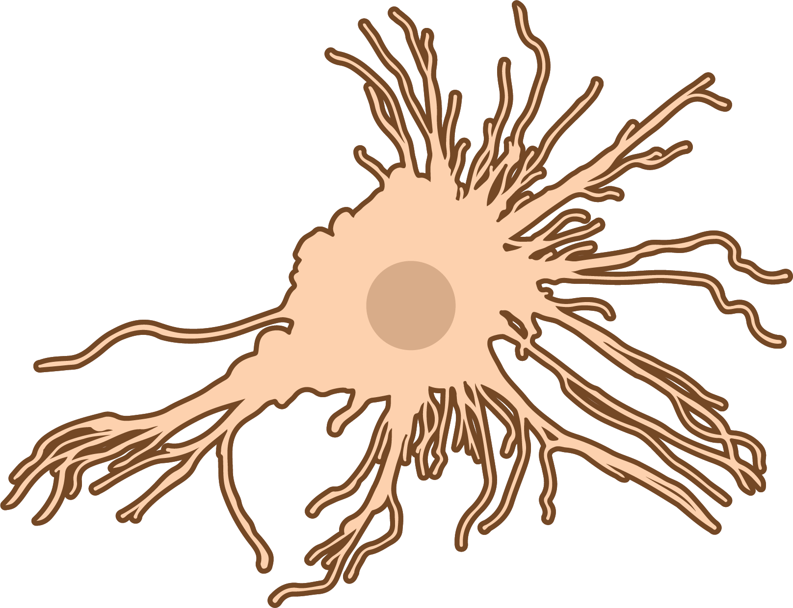
Peptide cancer vaccines are simple to make, but on their own they often fail to rouse a strong immune response. Several 2026 papers take on that problem in two ways. Mouse studies are testing nanoparticle carriers that deliver peptide antigens together with immune-activating partners, and reviews are looking at mutant calreticulin, a marker found only on blood-cancer cells, as a target for vaccines and other immune approaches [1, 2, 3, 4, 5]. Almost all of this work is preclinical or early-stage.
Key takeaways
- In mouse tumor models, lipid nanoparticles that carry HPV peptide antigens together with an immune-activating partner slowed tumor growth, with no obvious toxicity reported [3, 4]. These are animal results and may not carry over to people.
- A liposomal gastric cancer peptide vaccine made antibody and T-cell responses stronger and extended survival in mice [5].
- Two reviews describe mutant calreticulin as a tumor-specific neoantigen in myeloproliferative neoplasms. Peptide vaccines are one of several approaches under study, and an experimental antibody has the most mature clinical data so far [1, 2].
- Human data on peptide vaccines in this area remain limited. One registry entry, for a lung cancer peptide vaccine trial, lists a recruiting phase 1/2 study with no results posted [6].
Why peptide vaccines need help
A peptide vaccine uses short protein fragments (antigens) that are meant to train the immune system to recognize cancer cells. The difficulty is that short peptides on their own tend to provoke only a weak response. They can also struggle to reach the immune cells that need to process them [3, 5].
The gastric cancer study puts it plainly: its target antigen is promising, but “its clinical application is hampered by low immunogenicity” [5]. The HPV nanoparticle study likewise names “insufficient immunogenicity and inefficient intracellular delivery” as the main limits of therapeutic HPV peptide vaccines [3].
The usual fix is to pair the antigen with an adjuvant (an immune-activating partner) and to package both so they reach lymph nodes together. The studies below test different versions of that idea.
Nanoparticle carriers for HPV peptide antigens (mouse studies)
Phenolic lipid nanoparticles with manganese. One study built a lipid nanoparticle that holds HPV16 E6/E7 peptide antigens through hydrogen bonding rather than electrical charge [3]. The authors say this lets it carry peptides with a range of chemical properties [3]. The same particle also binds manganese ions, used here as an adjuvant [3].
In lab tests, the particles increased antigen uptake and helped dendritic cells mature. They also activated STING-related signaling, a pathway involved in innate immune alarms [3]. After injection into muscle, the particles reached draining lymph nodes and kept antigen there longer [3]. In the TC-1 model, a mouse tumor model of HPV-associated cancer, mice already carrying tumors showed more CD8+ T cells inside tumors, fewer immunosuppressive cells and slower tumor growth, with no systemic toxicity observed [3].
This is a cell and animal study, and the abstract does not give group sizes. “No observable toxicity” in mice is not a human safety finding.
Nanomicelles combining a peptide vaccine with chemotherapy. A second study paired tumor peptides (HPV16 E7, or the mouse neoantigen Adpgk) with a modified form of SN38, the active form of the chemotherapy drug irinotecan [4]. The components self-assembled into particles about 115 nm across, with more than 60% of the peptide captured [4].
In mouse models of HPV-positive TC-1 and MC38 colon tumors, a short course before surgery shrank primary tumors [4]. It also prevented recurrence after surgery in the treated groups and left long-lasting memory T cells [4]. Tumors showed more CD8+ T cells and fewer regulatory T cells and myeloid-derived suppressor cells, with no discernible systemic toxicity [4].
The authors call the platform “clinically translatable” [4]. That is a hope, not a demonstrated result: the evidence is from mice only, and the abstract does not report animal numbers.
A carrier-plus-helper design for gastric cancer (mouse study)
A third study targeted MG7-Ag, an antigen that appears abnormally in several gastrointestinal cancers [5]. The researchers built a trivalent peptide (three repeats of one fragment) and chemically linked it to a T-helper epitope taken from diphtheria toxin [5]. They placed it in liposomes with α-galactosylceramide, a molecule that activates natural killer T cells [5].
In mice, this combination produced higher levels of specific IgG antibodies than the linked peptide alone or the unlinked peptide with α-galactosylceramide [5]. It also increased IFN-γ and IL-4 release and CD4+/CD8+ T-cell responses [5]. The antibodies bound tumor cells and triggered complement-mediated killing [5]. In a mouse ascites tumor model, vaccinated animals built up less fluid and survived longer [5].
This is an animal study with no n given in the abstract. The ascites model is a fast-growing mouse system and is not a direct model of human gastric cancer.
Mutant calreticulin: a marker only cancer cells carry
The second fix is about the target itself. A good vaccine target sits on cancer cells but not on healthy ones. Mutant calreticulin may come close to that ideal in myeloproliferative neoplasms (MPNs), a group of chronic blood cancers [1, 2].
Calreticulin is a normal protein that helps cells fold other proteins and supports immune signaling [1]. Mutations in the CALR gene change its tail. The altered protein escapes its usual home in the endoplasmic reticulum, binds the thrombopoietin receptor and switches on growth signaling [1, 2]. These mutations account for about 30-35% of myelofibrosis cases, the most severe form of MPN [2]. Because the mutant tail creates new protein sequences, called neoepitopes, it is a tumor-specific neoantigen absent from normal tissue [1, 2].
Both papers are reviews, not new trials. They describe a range of approaches aimed at mutant calreticulin: peptide vaccines, monoclonal antibodies, bispecific T-cell engagers, antibody-drug conjugates and CAR T-cell therapy [1, 2]. According to the reviews, the most clinical data so far come from an experimental Fc-silenced monoclonal antibody, not from a vaccine [1, 2]. Early reports describe improvements in spleen size, symptoms and anemia, along with early markers of disease modification [2]. One review notes emerging evidence that the antibody may shrink the mutant cell population while sparing normal blood cell production [1].
The reviewers are cautious. They say the durability and clinical significance of these molecular responses are uncertain [1]. They also list immune evasion and differences between mutation subtypes as open challenges [1]. One review discusses how these approaches might eventually change decisions about donor stem-cell transplant, which is currently limited to patients fit enough and with a suitable donor [2]. That discussion is forward-looking, not based on completed trials.
Peptide vaccines in people: an early signal from the registry
Human evidence for the carrier designs above does not yet exist; all three were tested in animals [3, 4, 5]. Separately, a registry entry describes a phase 1/2 study of a MUC1 peptide vaccine in non-small cell lung cancer, focused on immune response [6]. Its status is listed as recruiting, with the record last updated in October 2026 [6]. A registry record describes a study plan, not its results, so it says nothing yet about whether the vaccine works.
What we still don’t know
- Whether mouse results carry over to people. The nanoparticle and liposome findings come from mouse tumor models [3, 4, 5]. Tumor control and low toxicity in mice often do not translate to humans.
- How large the studies were. None of the abstracts for the animal studies report group sizes, so the strength of their findings is hard to judge [3, 4, 5].
- How peptide vaccines compare with other ways of targeting mutant calreticulin. The reviews cover vaccines alongside antibodies and cell therapies, but the clinical data highlighted so far concern an antibody [1, 2].
- Whether early molecular responses last or matter clinically. The reviewers flag durability, immune evasion and mutation-subtype differences as unresolved [1].
- Results from human peptide vaccine trials. The lung cancer trial in the registry has no posted results [6].
This article is based on the published abstracts of the cited papers and on a trial registry record.
References
- Calreticulin and myeloproliferative neoplasms: linking biology to targeted therapies. Expert Review of Clinical Immunology (2026). doi:10.1080/1744666X.2026.2749708
- How could calreticulin-targeting therapies change the myelofibrosis transplant paradigm?. Bone Marrow Transplantation (2026). doi:10.1038/s41409-026-03060-3
- Phenolic Hydroxyl-Containing Lipid Nanoparticles Enable Codelivery of HPV Peptide Antigens and Manganese Adjuvant for Cancer Immunotherapy. ACS Applied Bio Materials (2026). doi:10.1021/acsabm.6c01833
- The lipid nanomicelles incorporating peptide vaccine and SN38 prodrug potentiate neoadjuvant chemo-immunotherapy. International Journal of Pharmaceutics: X (2026). doi:10.1016/j.ijpx.2026.100681
- A trivalent gastric cancer antigen conjugated to a diphtheria toxin epitope synergizes with an NKT cell agonist to boost antitumor immunity. Frontiers in Immunology (2026). doi:10.3389/fimmu.2026.1903458
- Study of the Immune Response of MUC1 (Mucin1) Peptide Vaccine for Non-small Cell Lung Cancer. ClinicalTrials.gov NCT01720836, status: recruiting.
Image credits
Free to use (CC0 / public domain); credited as a courtesy.
- Dentritic Cell (NIH BioArt 116 – 629005).png, by Courtesy of NIAID
Ryan Kissinger (Public domain, via wikimedia-commons): https://commons.wikimedia.org/wiki/File:Dentritic_Cell_(NIH_BioArt_116_-_629005).png
– Bone marrow.png, by Yuki T. Okamura (CC0, via wikimedia-commons): https://commons.wikimedia.org/wiki/File:Bone_marrow.png